Orthopaedics
Understanding elbow dysplasia
Lameness in a front leg, stiffness after rest or reluctance to exercise can point to elbow dysplasia. The term does not describe a single disease but several developmental abnormalities of the joint, which may occur on their own or together.
What is elbow dysplasia?
The elbow is a complex joint formed by three bones: the humerus, the radius and the ulna. To work normally, their surfaces must fit together correctly.
Elbow dysplasia is not a single disease. It groups together several developmental abnormalities, including:
- fragmented medial coronoid process, sometimes with a fissure or a bone fragment;
- ununited anconeal process, when this part of the ulna does not fuse normally;
- osteochondrosis or osteochondritis dissecans (also seen in the shoulder), which affects the joint cartilage;
- incongruity, when the bones do not fit perfectly within the joint.
Several of these abnormalities can coexist. They can cause pain and inflammation, and then lead to osteoarthritis.[1, 2]
The condition is seen more often in medium and large dogs, but other dogs can be affected too. Hereditary and environmental factors may both play a part; the exact cause is not the same in every case.[1]
Which signs should raise concern?
Signs often appear in young dogs, but some animals are only diagnosed later, once osteoarthritis or pain becomes more obvious.
You may notice:
- lameness in a front leg, sometimes intermittent at first;
- lameness that gets worse after exercise or does not fully go away with rest;
- stiffness after a period of rest;
- reluctance to walk for long or to play;
- discomfort when the elbow is bent or stretched.
Both elbows can be affected. In that case the lameness may be less obvious, because both legs are uncomfortable. The dog may mainly seem to have less stamina or be less keen to move around.[1]
These signs alone are not enough to conclude that a dog has elbow dysplasia. Other problems can cause front-leg lameness: a veterinary examination is needed to find the cause.
How is the diagnosis made?
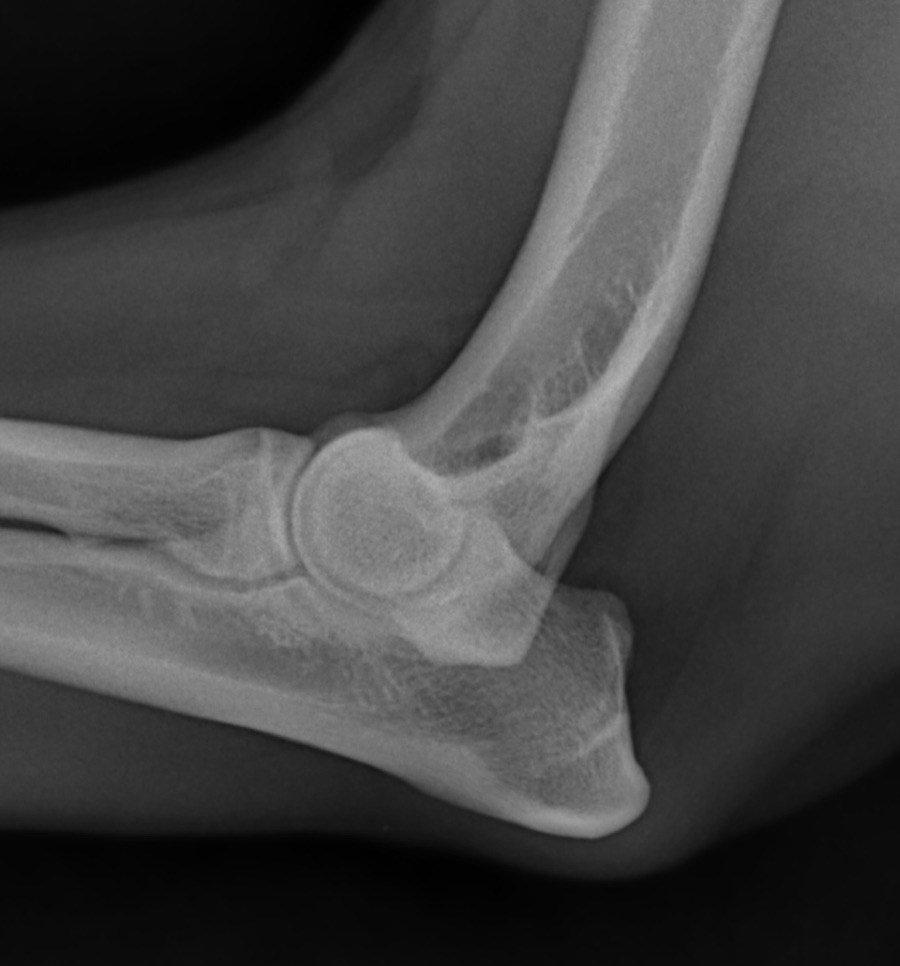
The vet watches your dog walk, examines the legs and manipulates the elbows. X-rays are usually taken to look for bone abnormalities or signs of osteoarthritis.
However, X-rays do not always answer every question, especially when a problem with the medial coronoid process is suspected. Depending on the examination and the images obtained, a CT scan or an arthroscopy may be suggested to look at the joint more precisely.[1, 3]
The choice of tests therefore depends on each dog: its signs, the clinical examination and what the first images show.
What treatments are available?
Treatment depends on the abnormality present, how severe it is and the condition of the joint. It may combine medical treatment, adjustments to activity and, in some cases, surgery.
Arthroscopy — a procedure performed with a small camera inserted into the joint — may be considered for some lesions. Other situations may call for a different technique, or for medical management. The decision is made case by case.[1]
Once osteoarthritis is established, treatment aims to limit pain and preserve comfort and mobility. Surgery can treat some lesions, but it does not remove osteoarthritis that is already present, which may continue to progress. Long-term follow-up is therefore sometimes needed.[1, 2]
What is the outlook?
The prognosis depends on the lesion, how it develops and how much osteoarthritis is already present. An accurate diagnosis helps choose the most suitable treatment and set realistic goals.
Even when osteoarthritis progresses on X-rays, comfort and lameness can improve with appropriate treatment. Progress nonetheless varies from one dog to another.[1]
When should you see a vet?
Book an appointment if your dog is limping on a front leg, seems stiff after rest or is gradually cutting back on walks and play. Lameness that comes back or persists deserves an examination, even if it is better on some days.
Key points
Elbow dysplasia covers several abnormalities that can look alike and sometimes coexist. An orthopaedic examination and imaging allow the vet to pin down the lesion and discuss the treatment options.
Care at Colvet
If your dog is limping or elbow dysplasia is suspected, your regular vet can examine your dog and refer you for an orthopaedic work-up if needed. Colvet also sees referred dogs for veterinary orthopaedic examinations and opinions, in Luxembourg.
Scientific sources
This article is based on the following veterinary publications:
- American College of Veterinary Surgeons (ACVS). Canine Elbow Dysplasia. Abnormalities involved, clinical signs, diagnosis, treatment and prognosis. acvs.org/small-animal/canine-elbow-dysplasia
- Elbow Dysplasia in Dogs. MSD Veterinary Manual, chapter “Arthropathies and Related Disorders in Small Animals”. msdvetmanual.com
- Villamonte-Chevalier A, van Bree H, Broeckx BJG, Dingemanse W, Soler M, Van Ryssen B, Gielen I. Assessment of medial coronoid disease in 180 canine lame elbow joints: a sensitivity and specificity comparison of radiographic, computed tomographic and arthroscopic findings. BMC Veterinary Research. 2015;11:243. doi.org/10.1186/s12917-015-0556-9