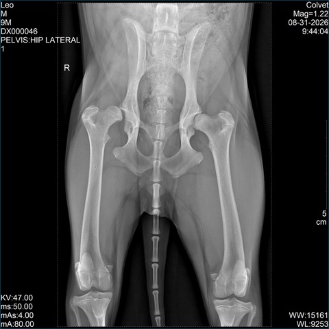
Hip dysplasia is common in medium and large breed dogs, yet it can go unnoticed for a long time. Some dogs run, play and never limp, even though their hips are already abnormal.
The absence of lameness does not mean the joint is healthy. When a hip lacks stability, the abnormal forces applied to the cartilage can gradually lead to osteoarthritis. Identifying the problem early therefore makes it possible to put suitable measures in place before the damage becomes significant.
What is hip dysplasia?
The hip is formed by the head of the femur, normally held within a socket in the pelvis called the acetabulum. In a dysplastic dog, the joint develops abnormally and shows excessive laxity: the femoral head is not stable enough within the acetabulum.
With every movement, this poor joint congruence creates abnormal stresses. Over time, they can deform the joint and lead to osteoarthritis.
Dysplasia is a complex, multifactorial disease. Genetic predisposition plays a major role, but growth, nutrition, body weight and certain physical stresses can influence how it is expressed and how it progresses.
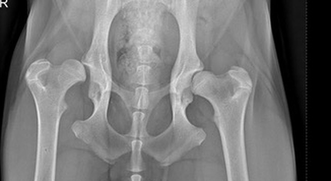
Close-up on both hips: what an X-ray shows is how the femoral head sits within the acetabulum, and how much contact there is between the two.
Which dogs can be affected?
Dysplasia is more often seen in certain medium, large or giant breeds, in particular the German Shepherd, the Labrador, the Golden Retriever, the Rottweiler, the Bernese Mountain Dog and the Newfoundland.
It can nevertheless affect a dog of any breed, including a crossbreed or a smaller dog. Nor does the absence of a known history in the parents completely rule out the risk.
Which signs should you watch for?
Signs vary with the dog's age and with how far the disease has progressed. They may include:
a swaying gait of the hindquarters;
a "bunny hopping" run, with both hind limbs moving together;
reluctance to run, jump or climb stairs;
difficulty getting up after resting;
reduced stamina, or a tendency to cut walks short;
In a young dog, the signs may be intermittent and can sometimes disappear for a while. In an adult dog, the discomfort is more often linked to secondary osteoarthritis.
Above all, some dysplastic dogs show almost no signs at all. Their adaptation, their muscle mass and their individual pain tolerance can mask the problem. Watching the way a dog moves is therefore not always enough to rule dysplasia out.
Why is an early diagnosis useful?
Screening is not only about giving the hips a "grade". It answers several practical questions:
are the joints stable?
are there already signs of osteoarthritis?
do the hips really explain the symptoms observed?
should the dog's weight, activity or follow-up be adjusted?
is a joint-preserving surgical option still feasible?
In some predisposed puppies, laxity can be assessed before the age required for official radiographic screening, which the Fédération Cynologique Internationale sets at twelve months for most breeds and eighteen months for large and giant breeds. This distinction matters: waiting for adulthood to obtain an official grading is not always the same thing as waiting to look for an abnormality in an at-risk or symptomatic dog.
Some procedures intended to improve the coverage and stability of the hip can only be offered during a limited growth window and before significant osteoarthritis has set in. A late diagnosis can therefore reduce the treatment options available.
How is hip dysplasia diagnosed?
The diagnosis starts with an orthopaedic consultation. The vet watches the dog move, looks for pain or a reduced range of motion in the hips, assesses muscle mass and checks that another condition — of the stifle or the spine, for instance — is not responsible for the symptoms.
X-rays are then taken. Very precise positioning and sufficient muscle relaxation are essential to interpret the hips correctly, so sedation or a short general anaesthetic is usually necessary.
What do the X-rays show?
Depending on the dog's age and on the purpose of the examination, different views or methods may be used. X-rays notably allow assessment of:
the congruence between the femoral head and the acetabulum;
the coverage of the femoral head;
the shape of the different parts of the joint;
the presence and severity of osteoarthritis.
The findings must always be interpreted alongside the clinical examination. A very abnormal X-ray does not necessarily mean severe pain, while an uncomfortable dog may show only subtle radiographic changes. We treat a dog, not just an image.
When the dog has few or no symptoms
Management depends on age, weight, activity level, joint laxity, the osteoarthritis already present and, above all, on how much the dog is bothered.
Finding dysplasia does not mean surgery is automatically needed. Monitoring and preventive measures may be all that is indicated:
maintaining an optimal body weight;
encouraging regular, progressive activity;
avoiding sudden changes in exercise;
maintaining hind limb muscle mass;
adapting the activities that trigger pain;
arranging follow-up checks according to age and clinical progression.
Weight control is particularly important. Excess weight increases the loads applied to the hips and can worsen the consequences of osteoarthritis.
When the dog is painful
Treatment may combine, depending on the case, adjusted activity, weight loss, physiotherapy, pain relief or anti-inflammatory medication and overall management of osteoarthritis.
The aim is to preserve comfortable activity and a good quality of life. Treatment should be reassessed whenever the dog's pain, mobility or needs change.
When is surgery considered?
In some young dogs, joint-preserving surgery can be discussed when laxity is identified early enough and significant osteoarthritis has not yet developed.
When the joint is already severely damaged, or when pain remains insufficiently controlled, other procedures may be offered. A total hip replacement aims to replace the painful joint. In certain situations, excision of the femoral head and neck may also be considered.
There is therefore no single operation suited to every dysplastic dog. The choice depends on the stage of the disease, the patient's build, its lifestyle and the goals being pursued. These procedures are planned and carried out in our surgical suite.
Can hip dysplasia be prevented?
It is not always possible to prevent it, because the genetic component is significant. Several factors that influence how it is expressed and what it leads to can nevertheless be acted upon:
choosing a puppy from properly screened parents;
avoiding excessive weight gain during growth;
feeding a complete diet suited to the dog's size and age;
favouring regular, progressive and appropriate activity;
having a predisposed puppy, or one showing suspicious signs, examined early.
Screening breeding dogs remains essential to reduce the frequency of the disease over successive generations.
When should you book an appointment?
A consultation is recommended if your dog is limping, stiff, moving unusually, struggling to get up or losing physical ability.
Screening can also be worthwhile in the absence of any symptom:
in a puppy of a predisposed breed;
when a family history is known;
before a dog is used for breeding.
If your dog is limping and no cause has been found, our article on lameness with no visible cause explains how the work-up continues.
How we manage it at Colvet
At the ColVet practice in Cessange, the assessment includes an orthopaedic examination and, when indicated, X-rays taken in a suitable position. The aim is to establish whether the hips are genuinely responsible for the discomfort, and to offer management tailored to the dog's age, activity and stage of disease.
We describe our approach to this condition on the page dedicated to hip dysplasia, and all of our procedures on the orthopaedic surgery page.
Dysplasia found early does not mean an operation is required. Above all, it tells you where the joints stand and lets you act while the widest range of options is still open.
Scientific sources
This article draws on the following veterinary publications and documents:
Fédération Cynologique Internationale. FCI Requirements for Official Hip Dysplasia Screening. fci.be (PDF)
Fédération Cynologique Internationale. Radiographic Procedure for Hip Dysplasia Evaluation. fci.be (PDF)
Schachner ER, Lopez MJ. Diagnosis, prevention, and management of canine hip dysplasia: a review. Veterinary Medicine: Research and Reports. 2015;6:181–192. doi.org/10.2147/VMRR.S53266
Smith GK, Paster ER, Powers MY, Lawler DF, Biery DN, Shofer FS, McKelvie PJ, Kealy RD. Lifelong diet restriction and radiographic evidence of osteoarthritis of the hip joint in dogs. Journal of the American Veterinary Medical Association. 2006;229(5):690–693. doi.org/10.2460/javma.229.5.690
General information article. It does not replace a consultation: only a veterinary examination can establish a diagnosis and offer the treatment suited to your animal.
Unsure about your dog's hips?
Early screening keeps more options open
Orthopaedic consultation and hip X-rays at the practice in Cessange, both for predisposed puppies and for dogs that are already uncomfortable.