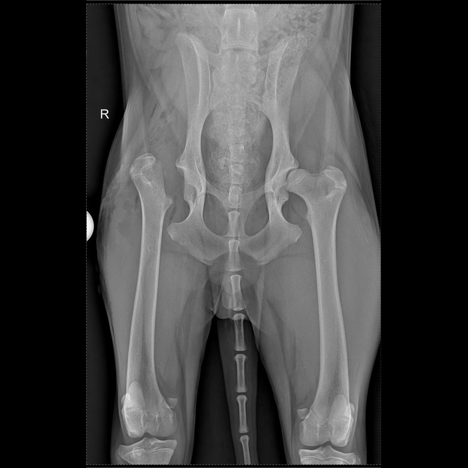
What is a hip luxation?
The hip is made up of the head of the femur, which normally sits in a socket of the pelvis called the acetabulum. It is held in place by the joint capsule, a ligament running through the centre of the joint and the muscles surrounding the pelvis.
During a luxation, the femoral head leaves the acetabulum completely. The structures that hold the joint in place are then stretched or torn.
In dogs, the femoral head most often moves upwards and forwards relative to the pelvis. This is called a craniodorsal luxation.
How does a hip become dislocated?
Luxation usually follows trauma:
- a road traffic accident;
- a fall;
- a violent impact;
- an accident while running or playing;
- a limb caught or trapped;
- a collision with another animal.
Because considerable force is needed to dislocate a healthy hip, other injuries may be present: fractures, chest or abdominal trauma, spinal injury or damage to another limb.
More rarely, a luxation can occur after relatively moderate trauma when the hip was already unstable, in particular when dysplasia is present.
What are the signs?
Signs usually appear suddenly. The dog may show:
- complete refusal to bear weight on the limb;
- marked pain;
- an abnormal position of the leg;
- a limb that appears shorter;
- difficulty or inability to stand up;
- reluctance to be touched around the hip;
- swelling or wounds linked to the trauma.
The position of the limb depends on the direction in which the femoral head has moved. Outward appearance alone therefore cannot confirm a luxation. Other causes of sudden lameness can look very similar.
What should you do straight away?
A hip luxation needs to be examined quickly. While waiting for the consultation:
- limit movement as much as possible;
- carry your dog if it can be handled safely;
- avoid slippery floors;
- do not pull on the limb;
- do not try to put the joint back in place yourself;
- do not give human painkillers.
After a major accident, the hip itself is not always the priority. Breathing, circulation and the search for internal injuries come first, before any orthopaedic manipulation or anaesthesia.
How is the diagnosis confirmed?
The vet starts by examining the whole dog, particularly when the luxation follows an accident.
The orthopaedic examination assesses the position of the limb, the pain, the mobility of the hip and any other injuries.
X-rays of the pelvis are essential. They make it possible:
- to confirm the luxation;
- to determine its direction;
- to look for a fracture of the pelvis or femur;
- to assess the shape of the femoral head and the acetabulum;
- to detect pre-existing dysplasia or osteoarthritis.
Further X-rays or investigations may be needed if multiple injuries are suspected.
Can the hip be put back in place without surgery?
Yes, in some situations. This manipulation is called a closed reduction: the femoral head is returned to the acetabulum without surgically opening the joint.
It is performed under general anaesthesia or, in some cases, under deep sedation combined with appropriate analgesia. The procedure would be far too painful in a conscious animal, and muscle contraction would prevent correct manipulation.
A closed reduction may be considered when:
- the luxation is recent;
- there is no joint fracture;
- the underlying shape of the hip is satisfactory;
- there is no significant osteoarthritis;
- the joint appears stable enough after reduction.
Success rates are generally better when the luxation is treated quickly, ideally within the first few days. Over time, muscle contraction, inflammation and the formation of tissue around the joint make reduction more difficult.
Why can the hip come out again?
Putting the femoral head back into the acetabulum does not immediately repair the structures that were torn during the accident.
A repeat luxation can be favoured by:
- a major tear of the joint capsule;
- poor stability after reduction;
- pre-existing dysplasia;
- a fracture that went unnoticed;
- tissue or fragments trapped inside the joint;
- a long-standing luxation;
- a sudden movement during convalescence.
Roughly one closed reduction in two stays in place in the long term. This estimate varies with the direction of the luxation, the state of the hip and the method of support used. Despite this risk of recurrence, closed reduction remains worthwhile when indicated, because it may avoid surgery (retrospective study of 171 dogs).
What happens after a closed reduction?
Follow-up X-rays are taken to check that the femoral head is correctly repositioned.
Depending on the direction of the luxation and the stability achieved, a bandage temporarily holding the limb in a set position may be proposed. This type of support is not suitable for every dog.
A hip bandage must be monitored very closely. It can slip, irritate the skin or cause pressure injuries. A study of craniodorsal luxations reported a high rate of complications with the Ehmer sling (Schlag et al., 2019). Its use must therefore be decided case by case and combined with frequent checks.
Strict activity restriction is then necessary. Outings are short and on the lead, with no running, jumping, stairs or play with other animals.
When is surgery needed?
Surgery may be indicated when:
- closed reduction is impossible;
- the hip comes out again immediately or quickly;
- the joint remains unstable;
- the luxation is long-standing;
- there is an associated fracture;
- the hip shows significant dysplasia or osteoarthritis;
- the shape of the joint makes a repeat luxation very likely.
Several techniques are possible. The choice depends on the age and weight of the dog, the state of the joint, any other injuries and the level of function being aimed for.
Surgical reduction and stabilisation of the hip
When the joint can be preserved, the hip is put back in place surgically and then stabilised.
Different techniques can be used:
- reconstruction or reinforcement of the joint capsule;
- placement of a prosthetic ligament of the femoral head;
- stabilisation with an anchor or a toggle pin type system;
- other constructs adapted to the direction of the luxation.
The implants hold the hip temporarily while the damaged tissues heal. The conformation of the joint must nevertheless be good enough to hope for lasting stability.
Femoral head and neck excision
When the hip cannot be kept in place in the long term, the head and neck of the femur can be removed. This procedure is also called femoral head and neck excision or resection.
A fibrous false joint gradually forms between the femur and the pelvis. The aim is to remove the painful bone-on-bone contact, not to rebuild an anatomically normal hip.
The outcome depends in particular on:
- the weight and build of the dog;
- its muscle mass;
- the extent of any pre-existing damage;
- the quality of the rehabilitation;
- the presence of other orthopaedic conditions.
This operation can provide very good comfort in carefully selected dogs, even though a slight difference in gait or range of motion may remain.
Total hip replacement
A total hip replacement replaces the femoral head and the acetabulum with implants. It recreates a true joint and generally offers the recovery closest to normal function.
It may be considered when a luxation is associated with dysplasia or significant osteoarthritis, or when optimal joint function is the goal.
This surgery requires careful patient selection and strict adherence to the postoperative instructions.
What recovery should you expect?
Recovery time varies with the treatment carried out.
After a reduction or a surgical stabilisation, several weeks are needed for the structures around the hip to heal. Activity restriction of about six to eight weeks is frequently recommended, after which exercise is reintroduced gradually.
After a femoral head and neck excision, controlled mobilisation and rehabilitation are particularly important. They help preserve range of motion and encourage the formation of a functional false joint.
In every case the programme must be tailored to the individual. A dog that has been through a major accident does not recover in the same way as a dog with an isolated luxation.
What is the prognosis?
The prognosis is generally good when the luxation is treated quickly and associated injuries are correctly identified.
The main complications are:
- a repeat luxation;
- irritation caused by the bandage;
- infection or an implant problem after surgery;
- a lasting reduction in hip range of motion;
- muscle wastage;
- the gradual development of osteoarthritis.
A recurrence after closed reduction does not mean that nothing more can be done. Surgical stabilisation or another procedure can then be considered.