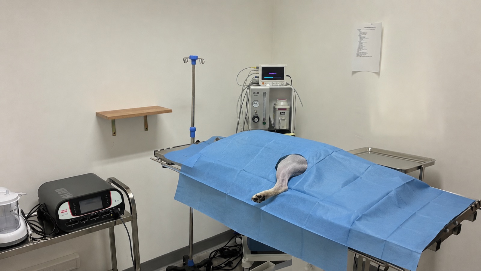
At Colvet, fluoroscopy lets us see the bone and the implants on a screen during surgery. For some fractures, this means we can realign the bone and stabilise it through small incisions, while leaving in place the muscles and small blood vessels around the break. Preserving these tissues can limit pain after surgery and support bone healing.[1]
Seeing through the skin during surgery
Whether the fracture is simple or complex, the main goal of osteosynthesis is to restore the broken bone's length and orientation. With fluoroscopy, this can be achieved without making a large incision in the patient's skin, because we can see the bone in real time through the soft tissues.[1, 2]
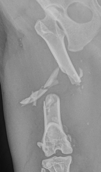
A femur fracture with several loose fragments.
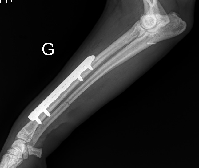
Through a small incision, the surgeon can slide the plate between the bone and the tissues around it. Fluoroscopy then lets them check the angle the plate needs and its length. The plate then bridges the fracture, where any small fragments stay attached to the tissues that nourish them.[1, 2]
A plate fixed on either side of the fracture.
Less tissue trauma, so less inflammation after surgery
Much of the pain after surgery comes from the inflammation caused by the operation itself. The skin has to be cut, and some tissues retracted or lifted away, to reach the bone, see the fracture and then repair it. By guiding the procedure through small openings, fluoroscopy helps limit this handling. The muscles and the sensitive membrane covering the bone suffer less additional trauma.[1]
Sparing these tissues reduces the inflammatory reaction caused by surgery and, with it, the pain felt in the first few days after the operation.[1]
Preserving small blood vessels and the haematoma can speed up healing
A fracture often creates loose fragments, completely separated from the rest of the bone. These fragments can only receive a blood supply through the tissues around them, via small blood vessels. In addition, in the first few days after a fracture, a haematoma forms around it and contributes to bone healing. The main aim of minimally invasive fixation techniques is to keep these structures as intact as possible. This preserves optimal conditions for the fastest and least painful recovery possible.[1]
A study of 16 dogs operated on for a tibial fracture illustrates this possible benefit. At 30 days, 5 of the 8 dogs treated with a minimally invasive plate had reached the stage of clinical union defined by the authors, compared with 2 of 8 after open plating. This sample is too small to predict how many days a given animal will gain.[1, 3]
How we handle this at Colvet
At Colvet, the choice of technique depends on the fracture. Some fractures are not well suited to minimally invasive techniques. But whenever we can limit your pet's inflammation and pain while speeding up bone healing, we think it is essential to do everything we can to make that possible.[1, 2]
Scientific sources
This article draws on the following veterinary publications:
Guiot and Déjardin (2011). Prospective evaluation of minimally invasive plating for 36 tibial fractures in dogs and cats. PMID 21223316
Boero Baroncelli et al. (2012). Comparison of healing after minimally invasive or open plating in 16 dogs. PMID 22828876
General information article. It does not replace a consultation: only a veterinary examination can establish a diagnosis and offer the treatment suited to your animal.
Has your pet broken a bone?
Every fracture is assessed before we choose the technique
X-rays on site and fluoroscopy in theatre: we explain which repair suits your pet, and why.