Cruciate ligament rupture in dogs : the signs on examination and on X-rays
A cruciate ligament rupture can be suspected without seeing the ligament itself. On examination, certain thickenings and abnormal movements of the knee are telling. On X-rays, the vet mainly looks for joint effusion, new bone formation and sometimes a forward shift of the tibia.
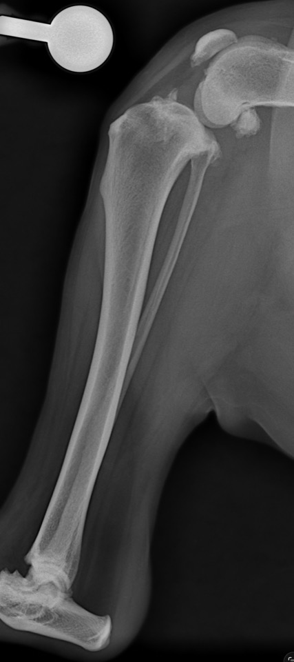
Side-view X-ray of a dog's knee taken at the practice.
Orthopaedics
The ligament can't be seen, but its consequences can
Diagnosing a cruciate ligament rupture relies on a body of evidence: what the vet feels under their fingers, abnormal movements of the knee and the changes visible on X-rays. Here are the practical signs to know.
How is a cruciate ligament rupture suspected?
The cranial cruciate ligament sits inside the knee (stifle). When it tears partially or completely, the joint becomes inflamed and unstable. The diagnosis rests on the agreement between abnormalities found on palpation, stability tests and the signs visible on X-rays.
The ligament itself is not visible on a standard X-ray. The images show its consequences on the joint, while the examination looks directly for thickening or abnormal movement of the knee.
On examination: a thickened patellar tendon
In a healthy dog, both edges of the patellar tendon — more accurately called the patellar ligament — are easy to feel, with a slight hollow on each side. When effusion and periarticular fibrosis develop, its outline becomes less distinct and the hollows disappear. The patellar ligament then feels as if it were buried in the infrapatellar fat pad.[6]
Comparing both knees at the same time helps reveal a difference that can be subtle.
On examination: the “medial buttress”
The medial buttress is a firm thickening that can be felt on the inner side of the knee, near the top of the tibia. It corresponds to periarticular fibrosis that develops in response to chronic instability.[6]
It is mostly found when the problem has been present for some time. It is easier to detect by comparing both knees with the dog standing.
On examination: cranial drawer and tibial thrust
The most direct sign is abnormal forward movement of the tibia relative to the femur. The vet can look for it in two ways:
with the cranial drawer test, moving the tibia forward while holding the femur still;
with the tibial thrust test, which reproduces the forces acting on the knee during weight-bearing.[6]
A clear movement confirms instability. It can, however, be hard to feel with a partial tear, in a very tense dog or when extensive fibrosis limits movement. The absence of an obvious drawer is therefore not enough to rule out a cruciate ligament problem.
Not all of these findings are specific, but together they strongly guide the rest of the work-up. They match the symptoms seen at home: hind-leg lameness, relapse once anti-inflammatories are stopped, difficulty sitting.
joint effusion and its effects on the soft tissues of the knee;
forward (cranial) displacement of the tibia relative to the femur, when present.
These changes must always be interpreted together with the orthopaedic examination.[1–4]
New bone formation: the most obvious chronic sign
A cruciate ligament rupture makes the knee unstable. Over time, this instability promotes osteoarthritis and the formation of new bone around the joint. These bony outgrowths are called osteophytes when they develop at the edges of the joint and enthesophytes when they appear where a tendon or ligament attaches.
They are often the easiest abnormalities to show on an X-ray of a long-standing or advanced case. Among the different signs of osteoarthritis, osteophytes are also one of the changes that progress most clearly over time.[1]
Published radiographic scoring systems assess the following sites in particular:
the upper and lower edges of the kneecap (patella);
the edges of the femoral trochlea;
the femoral condyles;
the cranial, caudal, medial and lateral margins of the tibial plateau;
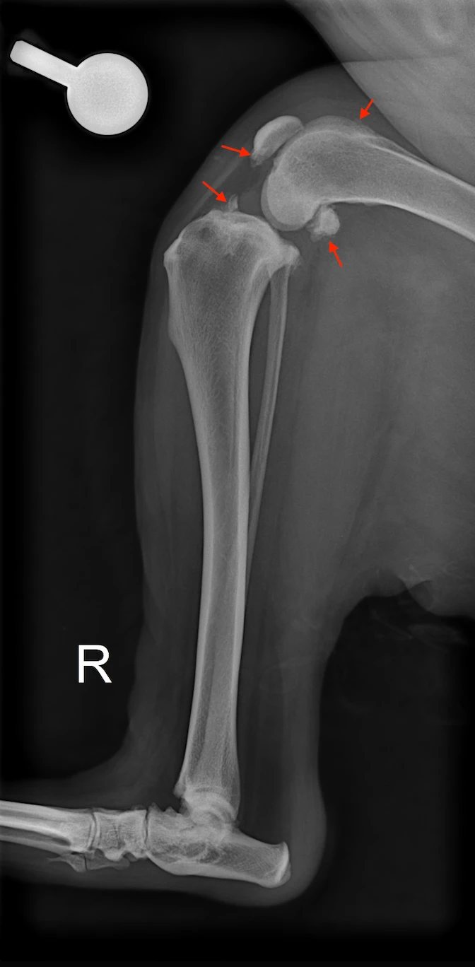
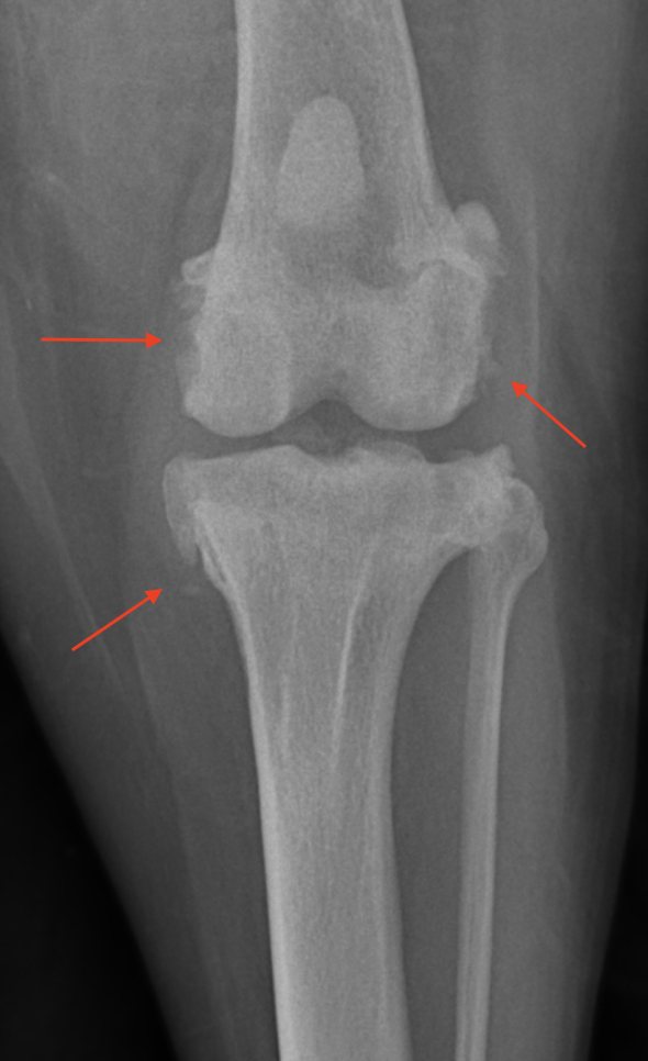
On a side view, new bone on the patella, trochlea, tibial plateau and fabellae is usually the most striking. On a front view, changes at the medial and lateral margins of the femur and tibia are easier to assess.
Left, side view: osteophytes on the patella, femoral trochlea, tibial plateau and fabellae. Right, front view: changes along the edges of the femur and tibia. This new bone reflects established osteoarthritis, frequently associated with the instability caused by cruciate ligament disease. Click to enlarge.
Joint effusion: often an earlier sign
When a knee is inflamed, synovial fluid builds up in the joint. The X-ray does not show this fluid distinctly, but it does show the changes it causes in the surrounding soft tissues.
On a normal side-view X-ray, the infrapatellar fat pad forms a darker triangular area just behind the patellar ligament. With effusion, this fat pad is compressed, pushed forward and becomes less clearly visible. The fat planes at the back of the knee may also be pushed backwards.[3, 4]
Loss of the infrapatellar fat pad outline is described as one of the early radiographic signs of cruciate ligament disease. It is nonetheless non-specific: any condition causing synovitis or effusion of the stifle can produce a similar image.[3, 4]
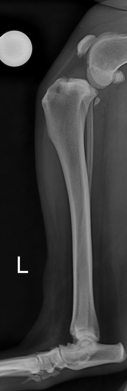
Left, a normal knee: the fat pad behind the patellar ligament forms a clearly defined dark area. Right, increased soft-tissue opacity within the joint compresses this fat pad. This appearance reflects joint effusion, common in cruciate ligament disease but not specific to it. Click to view the full X-rays.
Effusion and fat pad changes can also be present in the other knee before a clinically detectable rupture appears on that side.[3, 4] This is why both knees should be examined when cruciate ligament disease is suspected.
Forward shift of the tibia: a much more specific sign
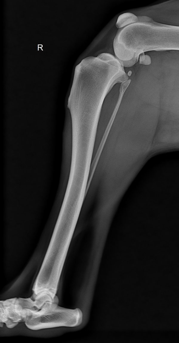
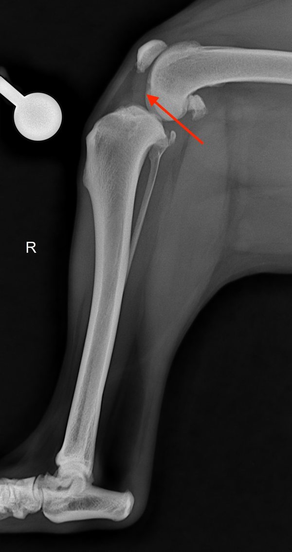
The cranial cruciate ligament normally limits forward movement of the tibia relative to the femur. After a complete rupture, the tibia may appear shifted forward on a side-view (mediolateral) X-ray.
When it is visible on a correctly positioned standard image, this abnormality is a very strong argument for a complete rupture.[5]
The tibia has shifted forward relative to the femur. This sign is strongly associated with a complete cruciate ligament rupture, but it is only visible in some affected dogs. Click to view the full X-ray.
This translation does not tell us whether the meniscus is injured: the meniscus has to be assessed separately.[5]
Other radiographic changes
Other abnormalities may accompany stifle osteoarthritis:
subchondral bone sclerosis;
thickening of the joint capsule and periarticular soft tissues;
thickening of the patellar ligament;
small areas of subchondral bone loss;
mineralisation within the joint or the meniscus.[1, 2]
These signs contribute to the overall assessment of the knee, but they point less directly to cruciate ligament disease than the combination of osteophytes, effusion and tibial instability. Subchondral sclerosis in particular is less reproducible between observers than scoring of osteophytes or effusion.[1]
What an X-ray cannot tell us
Even when it is very abnormal, an X-ray cannot:
show the cruciate ligament directly;
reliably distinguish a partial from a complete rupture when there is no subluxation;
confirm or rule out a meniscal tear;
measure precisely how much pain the dog is in.
The final diagnosis therefore rests on the agreement between the history, the orthopaedic examination — in particular the search for a cranial drawer or tibial thrust — and the radiographic signs. Sedation can help when pain or muscle tension prevents reliable handling and positioning.[6]
In practice: the signs that should raise suspicion
On examination
On the X-ray
Thickened patellar tendon with blurred edges
Loss of outline or forward displacement of the infrapatellar fat pad
Firm medial buttress
Osteophytes around the patella, trochlea, tibial plateau or fabellae
Cranial drawer or tibial thrust
Cranial tibial translation, when visible
Knee that is thickened, painful or less mobile
Capsular thickening and periarticular changes
Thigh muscle loss
Signs of osteoarthritis, more or less advanced
Several of these signs may be present at the same time, but no single picture is required. Partial tears in particular can cause effusion and a thickened patellar tendon without an easily detectable cranial drawer.
Not every sign is necessarily present in the same dog. Hind-leg lameness combined with new bone around the knee, effusion, a thickened patellar tendon or a medial buttress already warrants a further orthopaedic examination, even if no obvious cranial drawer could be demonstrated.
How we read X-rays at Colvet
At the Colvet practice in Cessange, X-ray interpretation always goes hand in hand with a full orthopaedic examination and, where appropriate, a comparison of both knees. We systematically look for new bone formation, signs of effusion and any forward shift of the tibia.
When the rupture is confirmed and TPLO is being considered, perfectly positioned X-rays are then used to measure the tibial plateau angle and plan the surgical correction. These procedures are performed in our surgical suite; our full approach to the cruciate ligament is described on our dedicated page.
Scientific sources
This article is based on the following veterinary publications:
Innes JF, Costello M, Barr FJ, Rudorf H, Barr ARS. Radiographic progression of osteoarthritis of the canine stifle joint: a prospective study. Veterinary Radiology & Ultrasound. 2004;45(2):143–148. doi.org/10.1111/j.1740-8261.2004.04024.x
Boyd DJ, Miller CW, Etue SM, Monteith G. Radiographic and functional evaluation of dogs at least 1 year after tibial plateau leveling osteotomy. Canadian Veterinary Journal. 2007;48(4):392–396. pubmed.ncbi.nlm.nih.gov/17494366
Fuller MC, Hayashi K, Bruecker KA, et al. Evaluation of the radiographic infrapatellar fat pad sign of the contralateral stifle joint as a risk factor for subsequent contralateral cranial cruciate ligament rupture in dogs with unilateral rupture. Journal of the American Veterinary Medical Association. 2014;244(3):328–338. doi.org/10.2460/javma.244.3.328
Chuang C, Ramaker MA, Kaur S, et al. Radiographic risk factors for contralateral rupture in dogs with unilateral cranial cruciate ligament rupture. PLOS ONE. 2014;9(9):e106389. doi.org/10.1371/journal.pone.0106389
Olson NJ, Weeren FR, van Eerde E. Correlation of spontaneous radiographic cranial tibial translation with complete cranial cruciate ligament rupture and medial meniscal tears in dogs. PLOS ONE. 2023;18(12):e0296252. doi.org/10.1371/journal.pone.0296252
Harasen G. Diagnosing rupture of the cranial cruciate ligament. Canadian Veterinary Journal. 2002;43(6):475–476. pubmed.ncbi.nlm.nih.gov/12058576
General information article. It does not replace a consultation: only a veterinary examination can establish a diagnosis and offer the treatment suited to your animal.